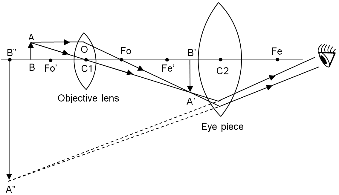
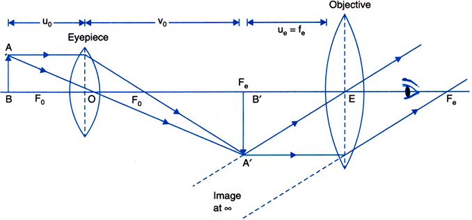
Ray compound resolving labelled
Table of Contents
Table of Contents
If you want to learn how to draw ray diagram of compound microscope, you’ve come to the right place. Drawing a ray diagram of a compound microscope can seem daunting at first, but with the right guidance, it can be a fun and rewarding experience.
When it comes to drawing a ray diagram of a compound microscope, there are a few common pain points that many people encounter. One of the biggest challenges is understanding the different components of the microscope and how they work together to create a clear image. Another challenge is figuring out how to properly align the lenses to create a sharp and detailed image.
To draw a ray diagram of a compound microscope, you will need to understand the basics of how the microscope works. A compound microscope is made up of two sets of lenses: the objective lens and the eyepiece. The objective lens is located at the bottom of the microscope and is used to magnify the specimen. The eyepiece is located at the top and is used to further magnify the image before it reaches your eye. To draw the ray diagram, you will need to determine the location and orientation of each of these lenses in relation to the specimen and your eye.
In summary, to draw a ray diagram of a compound microscope, you will need to understand the components of the microscope and how they work together, as well as how to properly align the lenses to create a clear image.
How to Draw Ray Diagram of Compound Microscope
If you’re ready to dive in and start drawing your own ray diagram of a compound microscope, here is a step-by-step guide to get you started:
Step 1: Start by drawing a simple diagram of the components of the microscope, including the lenses, the specimen, and your eye.
 Step 2: Next, determine the location of the objective lens and draw a line to represent the rays of light that are coming from the specimen.
 Step 3: Using the laws of refraction, draw the path of the rays through the objective lens.
 Step 4: Repeat this process for the eyepiece, drawing the path of the rays through the lens and into your eye.
 Tips and Tricks for Drawing a Ray Diagram of a Compound Microscope
Here are a few additional tips and tricks to help you create a clear and accurate ray diagram:
- Use a ruler to ensure that your lines are straight and accurate.
- Label each component of the microscope as you draw it to avoid confusion.
- Take your time and be patient. Drawing a ray diagram can take some practice, but it is a valuable skill to have.
Common Mistakes to Avoid When Drawing a Ray Diagram of a Compound Microscope
When you are first learning how to draw a ray diagram of a compound microscope, there are a few common mistakes that you may encounter. Here are a few things to watch out for:
- Forgetting to account for the eyepiece in your diagram.
- Failing to properly align the lenses.
- Overlooking small details, such as the thickness of the lenses.
Further Resources for Learning How to Draw a Ray Diagram of a Compound Microscope
If you are looking for more information on how to draw a ray diagram of a compound microscope, there are many resources available to help you. One great resource is online educational websites, which often have interactive tutorials and other learning materials. Additionally, textbooks and scientific journals can provide a wealth of information on the topic.
Question and Answer
Q: Why is it important to properly align the lenses when drawing a ray diagram?
A: Proper alignment of the lenses is crucial for creating a clear and accurate image. If the lenses are not aligned correctly, the image may be blurry or distorted.
Q: What is the purpose of the objective lens in a compound microscope?
A: The objective lens is used to magnify the specimen and create a clear image that can be viewed through the eyepiece.
Q: How can I tell if my ray diagram is accurate?
A: One way to check the accuracy of your ray diagram is to compare it to a diagram created by someone else who is experienced in drawing these types of diagrams. Additionally, you can double-check your work by referring to textbooks or scientific journals.
Q: How long does it take to learn how to draw a ray diagram of a compound microscope?
A: The amount of time it takes to learn how to draw a ray diagram can vary depending on your background knowledge and experience. With practice, most people can become proficient in drawing these diagrams within a few weeks or months.
Conclusion of How to Draw Ray Diagram of Compound Microscope
Drawing a ray diagram of a compound microscope can be a challenging but rewarding experience. By following the tips and tricks outlined above and practicing regularly, you can become proficient in creating clear and accurate diagrams. Remember to be patient and take your time, and don’t hesitate to seek out additional resources if you need help along the way.
Gallery
Draw A Labelled Ray Diagram To Show Image Formation By A Compound

Photo Credit by: bing.com / ray compound resolving labelled
Physics Compound Microscope Ray Diagram - Micropedia

Photo Credit by: bing.com / microscope
Compound Microscope | Fun Science

Photo Credit by: bing.com / microscope compound ray diagram objective eyepiece lens light working distance object draw simple construction diagrams explain physics correct eye inverted
Compound Microscope Derivation Class 12 - Micropedia

Photo Credit by: bing.com / compound microscope derivation magnification microscopes
A Compound Microscope Diagram - Micropedia

Photo Credit by: bing.com / microscope


